
X-ray Machine Safety: What You Need to Know
X-ray machines have revolutionized medical diagnostics, enabling healthcare providers to detect and assess a wide range of conditions. From broken bones to tumors, X-rays offer a non-invasive way to look inside the body. However, like any medical equipment, safety is paramount to ensure that the benefits of using X-rays far outweigh any risks associated with exposure to radiation. In this blog, we will explore the essential safety protocols for both healthcare professionals and patients when using X-ray machines.
BLOG
11/11/20243 min read

Understanding X-ray Radiation:
X-rays are a form of electromagnetic radiation, much like visible light but with much shorter wavelengths. This allows X-rays to pass through the body, capturing images of bones and soft tissues. The amount of radiation used in medical X-rays is typically low, but repeated exposure or high doses can pose health risks, including tissue damage or an increased risk of cancer. Therefore, understanding safety protocols is essential in preventing unnecessary exposure.
Safety Protocols for Healthcare Professionals:
For healthcare professionals operating X-ray machines, it is critical to follow strict safety guidelines to ensure their protection. Here are some key safety protocols:
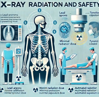
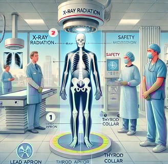
1. Use of Lead Shields:
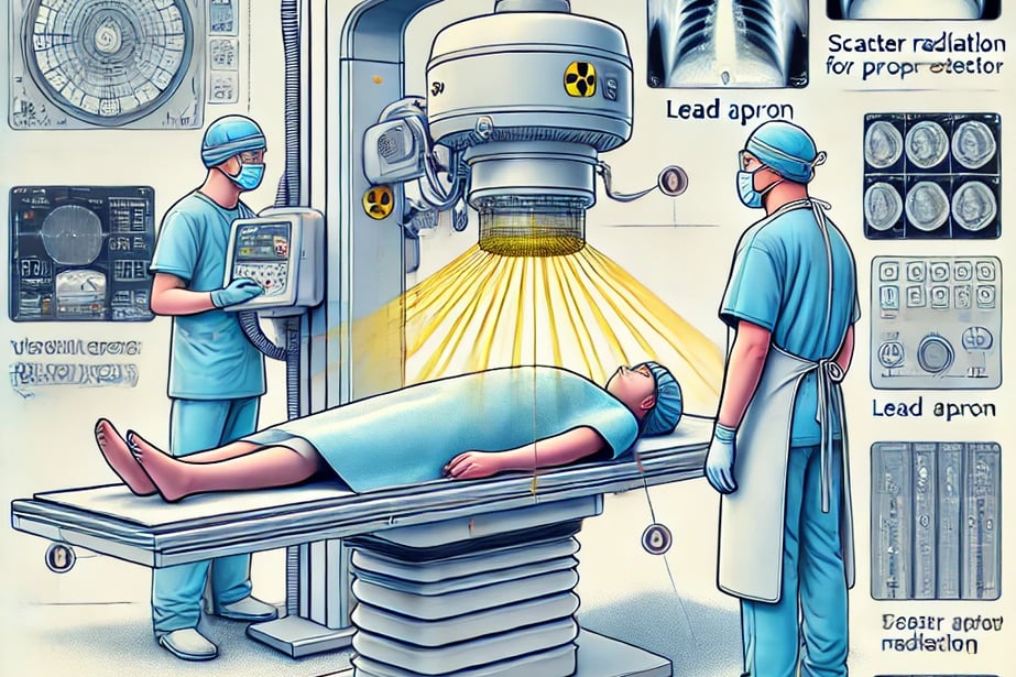
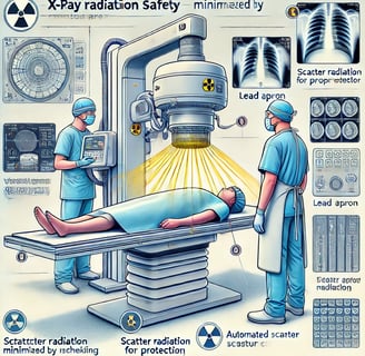
Lead aprons, thyroid collars, and lead gloves should be worn by both patients and operators when necessary to reduce exposure. These shields protect the body from scatter radiation, which can occur during an X-ray procedure.
2. Distance and Positioning:
Operators should always maintain a safe distance from the X-ray beam during imaging. Positioning is key—healthcare professionals should step away from the area of the X-ray and stand behind protective barriers or walls when the machine is in use.
3. Correct Settings on the Machine:
Modern X-ray machines come equipped with settings to ensure the lowest possible dose of radiation is used. Healthcare professionals should ensure the machine is set up correctly according to the patient’s needs, whether it’s a bone scan or a more complex imaging test.
4. Periodic Calibration and Maintenance:
Regular calibration and maintenance of the X-ray equipment are essential to ensure it is functioning properly. Poorly maintained equipment can increase radiation exposure unintentionally. Routine checks help reduce risks and enhance the overall quality of imaging.
Safety for Patients During an X-ray:
Patients also need to be informed and protected when undergoing an X-ray exam. Below are some important patient safety measures:
1. Pregnancy Considerations:
Pregnant women should inform their healthcare provider before undergoing an X-ray. In some cases, alternative imaging methods, such as ultrasound or MRI, may be considered to avoid the potential risks to the fetus.
2. Minimize Exposure:
X-rays should only be performed when necessary. Healthcare professionals should avoid redundant scans and ensure that each X-ray serves a clear diagnostic purpose.
3. Wearing Protective Gear:
Patients will often be asked to wear a lead apron during the X-ray, especially for areas of the body not being imaged, to reduce unnecessary exposure to radiation.
4. Proper Positioning:
Clear communication between the operator and patient is crucial to ensure correct positioning. This not only ensures the accuracy of the images but also limits the amount of radiation exposure by avoiding repeated X-ray images.
How Technology is Improving Safety in X-ray Procedures:
Advances in X-ray technology have dramatically improved safety for both healthcare professionals and patients. Some of the latest developments include:
1. Digital X-rays:
Digital X-rays use less radiation compared to traditional film-based X-rays and provide quicker, more accurate results. The images can be enhanced, reducing the need for additional exposures.
2. Automated Exposure Control:
Modern X-ray machines feature automated exposure control systems that adjust the radiation dose based on the patient’s size and the area being examined. This ensures the minimal necessary dose for high-quality images.
3. Three-Dimensional Imaging:
In certain cases, 3D X-ray imaging systems (like CT scans) allow for more detailed images without requiring multiple exposures, further reducing the patient’s radiation dose.
What to Expect During an X-ray Exam:
Here’s what you can expect when preparing for an X-ray:
Preparation: You'll be asked to remove any clothing or accessories that might interfere with the imaging (such as jewelry, glasses, or a bra with metal).
Positioning: The technician will guide you into the correct position to ensure the area being imaged is clearly visible.
The Scan: Once you are in position, the X-ray machine will emit a quick burst of radiation to capture an image. This typically takes only a few seconds.
Results: After the procedure, the images will be reviewed by a radiologist or the attending physician to diagnose your condition.

General Information about X-rays and Radiation:
Safety Guidelines for Healthcare Professionals:
Technological Advancements in X-ray Procedures:
Patient Information:
Cleveland Clinic - What to Expect During X-rays
Reference website link:
Innovative
Contact Us
Service
xraybazar.com
© 2024. All rights reserved.
Address- Rajasthan, India
Gmail Id- xraybazaroffcial.com
Important Links