
What is Radiology? Understanding the Role of X-Rays in Modern Medicine
In modern medicine, radiology plays an essential role in diagnosing and treating various health conditions. From identifying broken bones to detecting internal injuries or diseases like cancer, radiology and medical imaging have revolutionized how healthcare professionals understand the body and its functions. X-rays, a fundamental tool in the radiology field, have been pivotal in shaping medical practices for over a century. In this blog post, we will explore what radiology is, how X-rays work, and the vital role they play in modern healthcare.
1/31/20254 min read
1. What is Radiology?
Radiology is a branch of medicine that uses medical imaging techniques to diagnose and treat diseases within the body. The word "radiology" comes from radiation and ology (the study of). Radiologists—medical professionals specialized in interpreting medical images—use a variety of imaging methods to visualize the internal structures of the body and identify any potential abnormalities or diseases.
Radiology can be divided into two main categories:
Diagnostic Radiology: This involves using imaging technologies to view and diagnose conditions, including bone fractures, tumors, infections, or any other internal health issues.
Interventional Radiology: This involves minimally invasive procedures that are guided by imaging technologies. These procedures include biopsies, catheter placements, and even certain surgeries, all performed with the help of imaging tools like X-rays, CT scans, and ultrasound.
The goal of radiology is to provide detailed images that help doctors diagnose, plan treatment, and monitor the progress of conditions over time.
2. How Do X-Rays Work?
X-rays are a form of electromagnetic radiation that have high energy and the ability to penetrate through the body. The process of using X-rays in medical imaging is referred to as radiography.
Here’s how it works:
X-ray Machine: The patient is positioned in front of an X-ray machine, and the machine sends a beam of X-rays through the body.
Differential Absorption: As the X-rays pass through the body, they are absorbed by different tissues at different rates. Dense tissues, like bones, absorb more X-rays, while soft tissues (such as muscles and organs) absorb fewer.
Image Creation: The X-rays that pass through the body hit a special film or digital detector on the other side. The detector records the pattern of X-rays that pass through and converts it into an image.
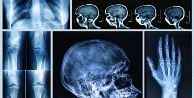
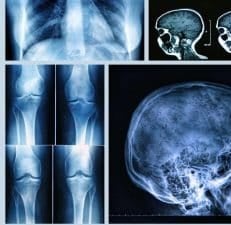
Interpretation: A radiologist examines the X-ray image to detect any abnormalities such as fractures, infections, tumors, or other health issues.
X-rays are incredibly useful because they provide clear images of bones and some soft tissues, making them valuable for detecting conditions like bone fractures, joint dislocations, pneumonia, and even cancer.
3. The Role of X-Rays in Modern Medicine
X-rays are one of the most commonly used and important tools in modern medicine. Here’s why they are so critical:
Diagnosing Fractures and Bone Disorders
X-rays are most commonly associated with orthopedic diagnostics. They are the go-to tool for identifying bone fractures, joint dislocations, arthritis, and conditions like osteoporosis. For example, when someone suffers a fall or injury, an X-ray can help quickly determine whether there is a fracture and how severe it is, which is crucial for providing the appropriate treatment.
Detecting Tumors and Cancer
X-rays are also used in the early detection of cancer. Mammography, a specialized type of X-ray, is widely used for breast cancer screening. It can detect small tumors long before they become palpable, improving the chances of early intervention and successful treatment. Similarly, X-rays and CT scans can be used to detect lung cancer, abdominal tumors, and cancers of the digestive system.
Assessing Chest Conditions
Chest X-rays are one of the most commonly performed medical imaging exams. They are used to diagnose a variety of chest-related conditions, including pneumonia, tuberculosis, lung cancer, pulmonary edema, and other lung diseases. A quick chest X-ray can provide critical information about a patient’s respiratory health and help physicians determine the best course of action for treatment.
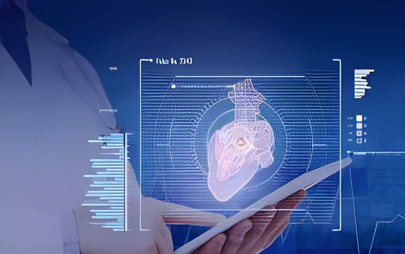
Monitoring Heart Conditions
While X-rays primarily provide images of bones and soft tissues, they can also help detect certain heart conditions. For example, an X-ray can reveal signs of heart enlargement, fluid buildup in the lungs (which may be a sign of heart failure), or cardiac tamponade (a buildup of fluid around the heart). These images can provide doctors with key insights into a patient's cardiovascular health.
Guiding Medical Procedures
Beyond diagnosis, X-rays also play a key role in interventional radiology. In this area, X-rays are used to guide medical procedures like the insertion of catheters, the removal of kidney stones, and even biopsy procedures. This ability to see inside the body in real-time allows doctors to perform surgeries with greater precision, reduce recovery times, and improve patient outcomes.
4. Advancements in X-Ray Technology
X-ray technology has come a long way since its discovery in 1895 by Wilhelm Roentgen. Today, we have several advanced imaging techniques that provide even more detailed and accurate results:
Digital X-Rays: Traditional X-rays used film to create images, but digital X-rays offer higher quality images with the added benefit of faster processing and lower radiation doses. Digital systems also allow images to be easily stored and shared, improving efficiency in healthcare facilities.
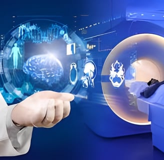
Computed Tomography (CT) Scans: CT scans, often referred to as “cat scans,” combine X-ray technology with computer processing to create detailed cross-sectional images of the body. CT scans can provide 3D images that offer a more comprehensive view of internal structures, making them invaluable for detecting and diagnosing a wide range of conditions, including cancers, internal bleeding, and infections.
Fluoroscopy: Fluoroscopy is a type of X-ray that allows doctors to observe real-time moving images of the inside of the body. It is used during certain medical procedures, such as placing a catheter or guiding a contrast agent through blood vessels to detect blockages.
3D Mammography: Traditional mammography produces flat, two-dimensional images of the breast, but 3D mammography, or tomosynthesis, creates three-dimensional images. This helps radiologists identify smaller tumors and differentiate between benign and malignant growths with greater accuracy.
5. Safety Considerations and Advances in Radiation Dose
One common concern with X-ray imaging is the potential risk of radiation exposure. While the amount of radiation used in most X-ray procedures is minimal, it is still important to minimize unnecessary exposure, especially for certain vulnerable groups such as pregnant women and young children.
Advances in X-ray technology have led to significant improvements in radiation safety. New equipment and techniques allow for lower doses of radiation to be used while maintaining image quality. For example, digital radiography allows for faster exposure times, reducing the total radiation dose. Additionally, radiologists are trained to use the lowest effective radiation dose necessary for obtaining clear, diagnostic images.

Reference Website Link:
American College of Radiology (ACR)
Link: https://www.acr.org
Radiological Society of North America (RSNA)
Link: https://www.rsna.org
National Institute of Biomedical Imaging and Bioengineering (NIBIB)
American Cancer Society - Radiation Therapy
Innovative
Contact Us
Service
xraybazar.com
© 2024. All rights reserved.
Address- Rajasthan, India
Gmail Id- xraybazaroffcial.com
Important Links