
How New X-Ray Machines Are Revolutionizing Diagnostic Imaging
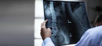
X-ray machines have been a cornerstone of medical diagnostics for over a century, providing doctors with essential insights into the internal structure of the human body. While traditional X-rays have played a vital role in detecting fractures, infections, and other medical conditions, the technology has come a long way since its invention. New innovations in X-ray machines are now changing the game, enabling faster, more accurate, and less invasive diagnostic imaging. In this blog post, we will explore how the latest advancements in X-ray machines are revolutionizing diagnostic imaging, improving patient care, and enhancing the overall healthcare experience.
3/10/20254 min read
1. High-Resolution Imaging: A New Era of Clarity
One of the most significant advancements in modern X-ray technology is high-resolution imaging. Traditional X-rays often offered clear but limited images, making it difficult to detect subtle or early-stage conditions. However, the latest X-ray machines provide exceptional clarity and detail, allowing healthcare providers to identify even the smallest fractures, tumors, or abnormalities.
With enhanced resolution, doctors can assess complex conditions more accurately, leading to better treatment planning and outcomes. For example, high-resolution X-rays can help dentists spot early signs of cavities, bone loss, or periodontal disease, while radiologists can more easily detect tumors or infections in soft tissues and organs.
Key Benefits:
Better accuracy in detecting conditions that may have been missed with older equipment.
Enhanced ability to detect early-stage conditions, which leads to quicker treatments.
Improved diagnostic capabilities for complex anatomical areas like the spine and joints.
2. Digital Radiography: Faster and More Efficient
The shift from traditional film-based X-rays to digital radiography (DR) has been one of the most transformative changes in the industry. Digital X-ray machines offer immediate, high-quality results, eliminating the need for film development and reducing the time patients spend waiting for their results.
With digital radiography, images are captured digitally and stored electronically, making it easier for healthcare providers to access, share, and analyze them. Radiologists can now view images in real-time, enabling them to make faster diagnoses and expedite treatment decisions.
Key Benefits:
Faster results, which improves workflow efficiency and reduces patient wait times.
Reduced radiation exposure compared to traditional film X-rays.
Easier storage and sharing of images for telemedicine or second opinions.
3. Portable X-Ray Machines: Increasing Accessibility
One of the most exciting developments in X-ray technology is the rise of portable X-ray machines. These compact devices are transforming how healthcare providers can offer diagnostic imaging, especially in emergency settings, critical care units, and remote locations.
Portable X-ray machines are ideal for patients who cannot move easily, such as those in intensive care or elderly patients in long-term care facilities. These machines can also be used in field hospitals or rural clinics, where access to traditional imaging equipment may be limited.
By bringing diagnostic imaging to the patient rather than the patient to the machine, portable X-rays improve patient comfort and ensure that timely diagnoses are made without delay.
Key Benefits:
Increased accessibility to diagnostic imaging, especially in underserved areas.
Reduced patient movement, which is crucial for those who are critically ill or immobile.
Faster response times for urgent medical conditions in emergency settings.
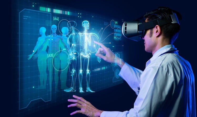
4. 3D X-Ray Imaging: A New Dimension in Diagnostic Precision
While traditional X-rays provide a flat, two-dimensional view of the body, 3D X-ray imaging (also known as cone beam CT or CBCT) adds a powerful new dimension to medical diagnostics. This technology captures multiple images from different angles and reconstructs them into a three-dimensional model, offering more detailed and precise views of the body’s internal structures.
In dentistry, orthopedics, and oncology, 3D X-ray imaging is proving invaluable for diagnosis, treatment planning, and surgical guidance. For example, in dental implants, 3D X-ray systems can accurately assess bone density and the precise positioning of teeth, reducing the risk of complications during surgery.
Key Benefits:
Enhanced visualization of bones, joints, and soft tissues for more accurate diagnosis.
Better treatment planning for complex cases like cancer or joint replacements.
The ability to detect problems that may be hidden in 2D images, such as small tumors or fractures.
5. Artificial Intelligence (AI) and Machine Learning in X-Rays
The integration of Artificial Intelligence (AI) and machine learning into modern X-ray systems is one of the most exciting breakthroughs in diagnostic imaging. AI can now be used to automatically analyze X-ray images, highlighting potential issues and assisting healthcare providers in making more accurate and timely diagnoses.
AI algorithms are trained to identify patterns in X-ray images that may be difficult for the human eye to detect, such as early-stage lung cancer, bone fractures, or pneumonia. By automating the initial analysis, AI reduces the risk of human error, speeds up the diagnostic process, and allows radiologists to focus on more complex cases.
Key Benefits:
Faster diagnosis through AI-assisted image analysis.
Increased accuracy, with AI helping to catch subtle conditions that may otherwise be missed.
Reduced workload for healthcare providers, allowing them to prioritize critical cases.
6. Lower Radiation Exposure: A Safer Approach
One of the main concerns with traditional X-rays is the exposure to ionizing radiation. Modern X-ray machines, however, are designed to reduce radiation doses while maintaining image quality. Advances in image processing and sensor technology allow for clearer images to be obtained with lower levels of radiation, making diagnostic imaging safer for patients.
Newer digital and 3D X-ray systems are particularly effective at reducing radiation exposure without sacrificing diagnostic clarity. This is especially important for pediatric patients, pregnant women, and anyone requiring repeated imaging.
Key Benefits:
Safer for patients, especially in sensitive populations like children and pregnant women.
Lower radiation levels while maintaining high-quality images.
Better patient safety, particularly for those who need frequent imaging.
7. Integration with Other Medical Technologies
Modern X-ray machines are increasingly integrated with other advanced medical technologies, such as electronic health records (EHRs) and robotic surgery systems. This integration allows for seamless sharing of X-ray images and diagnostic results across various healthcare platforms, making it easier for healthcare providers to collaborate and make informed decisions.
In addition, X-ray-guided surgery, often used in procedures like spine surgery or cardiac interventions, allows surgeons to monitor the progress of a procedure in real-time, improving precision and reducing the risk of complications.
Key Benefits:
Improved collaboration between different medical professionals.
Better surgical outcomes through real-time X-ray imaging during procedures.
Enhanced patient care with integrated technologies that provide comprehensive diagnostic information.


Reference Website Link:
RadiologyInfo.org (American College of Radiology) – https://www.radiologyinfo.org/
Siemens Healthineers – https://www.siemens-healthineers.com/
GE Healthcare – https://www.gehealthcare.com/
ScienceDirect - Diagnostic Imaging – https://www.sciencedirect.com/topics/medicine-and-dentistry/x-ray
Health Imaging News – https://www.healthimaging.com/
Innovative
Contact Us
Service
xraybazar.com
© 2024. All rights reserved.
Address- Rajasthan, India
Gmail Id- xraybazaroffcial.com
Important Links