
Exploring the Role of X-ray in Cancer Detection and Treatment
Cancer is a leading cause of death worldwide, but thanks to advancements in medical imaging, early detection, and treatment of various cancers have become more effective. One of the most essential tools in the fight against cancer is X-ray technology. X-ray imaging, including specialized techniques like mammography, CT scans, and fluoroscopy, plays a vital role in the detection, staging, and treatment of cancer. In this blog, we will explore how X-rays are used in cancer diagnosis and treatment, the benefits they offer, and the future possibilities for their role in cancer care.
3/4/20254 min read

1. X-rays for Early Cancer Detection
The earlier cancer is detected, the more treatable it becomes. X-ray technology is often the first line of defense in detecting certain types of cancer.
Mammography for Breast Cancer Detection
One of the most well-known uses of X-rays in cancer detection is mammography, a type of X-ray imaging used to detect breast cancer. Mammograms can identify lumps, changes in breast tissue, and abnormalities that may indicate the presence of cancer. Regular mammograms are critical for women, especially those over 40, as they can detect tumors before they are palpable, significantly improving treatment outcomes.
Benefits: Early detection of tumors, even in the earliest stages, allows for less invasive treatments and better chances for recovery.
Screening Guidelines: Regular screening helps identify cancers that are not yet causing symptoms, which can be crucial for survival rates.
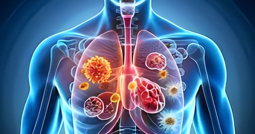
Chest X-rays for Lung Cancer Detection
Lung cancer is one of the deadliest cancers globally, but X-ray imaging plays a significant role in detecting it. Chest X-rays are often used to detect signs of lung cancer in patients who are at high risk (e.g., smokers or those with a family history). Though not as detailed as a CT scan, chest X-rays are a fast, cost-effective way to detect abnormalities in the lungs.
Benefits: X-rays help detect tumors, fluid accumulation, or other indicators of lung disease, which can then be further investigated with more advanced imaging techniques like CT scans.
X-rays in Bone Cancer
X-ray imaging is essential in diagnosing bone cancer (e.g., osteosarcoma). X-rays help visualize bone lesions or abnormalities, which can be crucial in determining the extent of cancer and planning further diagnostic or treatment steps.
Benefits: X-rays provide an initial overview of bone involvement, guiding doctors to pursue further tests, like MRI or CT scans, for more detailed imaging.
2. X-ray in Staging Cancer: CT Scans and PET Scans
Once cancer is detected, determining its stage—how far it has spread—is crucial for planning treatment. CT scans (computed tomography) and PET scans (positron emission tomography) are both forms of X-ray technology that provide detailed imaging to help in the staging process.
CT Scans for Cancer Staging
CT scans, which use multiple X-ray images to create cross-sectional images of the body, are instrumental in staging cancer. They can identify the size of tumors, whether they have spread to lymph nodes, or if they have infiltrated nearby organs.
Benefits: CT scans provide detailed, 3D images of tumors, helping doctors understand the cancer’s extent and plan surgical procedures or radiation therapy more accurately.
PET Scans for Detecting Metastasis
PET scans are sometimes combined with CT scans to create PET/CT imaging, offering detailed insights into both the structure and function of organs and tissues. This is particularly useful in metastatic cancer, where the cancer has spread to distant parts of the body.
Benefits: PET scans help doctors detect cancerous growths and determine whether the cancer has spread to other areas, enabling more precise treatment planning.
3. X-ray Technology in Cancer Treatment: Radiation Therapy
X-ray technology doesn’t just help detect cancer; it is also a key tool in treating it. Radiation therapy uses high doses of radiation, often in the form of X-rays, to target and destroy cancer cells. This non-invasive treatment can be effective in shrinking tumors, alleviating pain, or treating cancer that cannot be surgically removed.
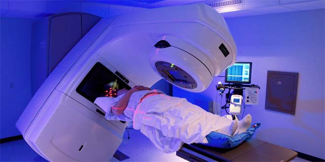
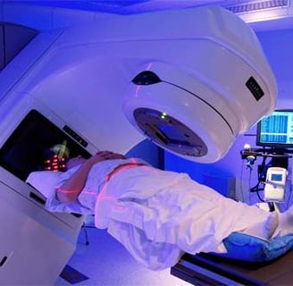
External Beam Radiation Therapy
In external beam radiation therapy, a machine directs high-energy X-rays precisely at the tumor. The X-rays damage the DNA of cancer cells, preventing them from growing and dividing.
Benefits: This method allows for targeted treatment, sparing nearby healthy tissue while focusing radiation on the tumor. It is commonly used to treat brain tumors, breast cancer, prostate cancer, and lung cancer.
Brachytherapy: Internal Radiation
In brachytherapy, radioactive sources are placed directly inside or near the tumor. This type of internal radiation therapy can be used to treat cancers in areas such as the prostate, cervix, and breast.
Benefits: It delivers a higher, more concentrated dose of radiation to the tumor while minimizing exposure to surrounding healthy tissue.
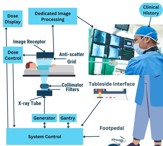
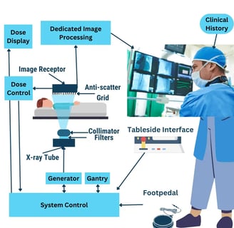
4. The Role of Fluoroscopy in Cancer Treatment Procedures
Fluoroscopy is a type of continuous X-ray imaging used to observe moving body parts in real-time. It is particularly useful in guiding various medical procedures used in cancer treatment.
Guiding Needle Biopsies
Fluoroscopy is used to guide needle biopsies when cancerous tissue needs to be sampled for analysis. By using live X-ray imaging, doctors can safely insert a needle into the tumor with great accuracy, even in hard-to-reach areas.
Benefits: It ensures precise targeting, reducing the risks associated with the procedure and increasing the likelihood of obtaining accurate tissue samples.
Radiation Treatment Planning
Fluoroscopy also assists in real-time monitoring of radiation therapy, ensuring that the radiation beams are directed precisely at the tumor, avoiding damage to surrounding healthy tissue.
Benefits: It improves the accuracy of radiation delivery, increasing the treatment’s effectiveness and reducing side effects.
5. Future of X-ray in Cancer Detection and Treatment
As technology continues to advance, the role of X-rays in cancer diagnosis and treatment will only expand. Here are some exciting developments to look out for:
AI in X-ray Imaging: Artificial intelligence is already enhancing the way doctors analyze X-ray images. AI algorithms can assist in detecting early-stage cancers, identifying patterns in X-ray images that might be difficult for the human eye to see.
Low-Dose X-rays: Innovations in X-ray technology are enabling the use of lower radiation doses for imaging, reducing the risks of exposure while maintaining diagnostic accuracy.
Enhanced 3D Imaging: 3D X-ray imaging offers more detailed, high-resolution views of cancerous tissues, making it easier to detect small tumors and plan targeted treatments more effectively.
Reference Website Link:
American Cancer Society (https://www.cancer.org/)
National Cancer Institute (https://www.cancer.gov/)
Radiological Society of North America (RSNA) (https://www.rsna.org/)
Mayo Clinic (https://www.mayoclinic.org/)
Cancer Research UK (https://www.cancerresearchuk.org/)
Innovative
Contact Us
Service
xraybazar.com
© 2024. All rights reserved.
Address- Rajasthan, India
Gmail Id- xraybazaroffcial.com
Important Links