
3D Imaging & Advanced Analysis: Revolutionizing X-ray Technology
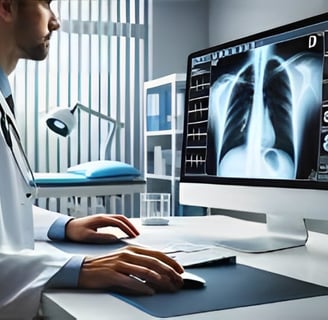
In recent years, 3D imaging and advanced analysis techniques have dramatically transformed the field of medical imaging. Traditionally, X-rays provided only 2D images, limiting the ability to assess complex structures or conditions. However, with the advancement of technology, 3D imaging has introduced new levels of precision, offering doctors, radiologists, and healthcare professionals powerful tools to enhance diagnostic accuracy and treatment planning. In this blog, we explore the role of 3D imaging and advanced analysis in modern X-ray technology and its profound impact on healthcare.
3/1/20255 min read

What Is 3D Imaging in X-rays?
Three-dimensional imaging in X-ray technology is a method in which several 2D X-ray images are obtained from different angles to build three-dimensional images of the inside of the body. The 2D images are processed and combined with the assistance of computer algorithms to build a three-dimensional image of the structures being studied.
In contrast to the conventional 2D X-rays, which can only take a two-dimensional picture, 3D imaging shows more and actual bones, organs, and soft tissues. 3D imaging is particularly beneficial in complicated medical conditions that need a general overview of the inner anatomy, including tumors, fractures, joint diseases, and vascular diseases.
How Does 3D Imaging Work?
3D image creation from routine X-rays generally has the following procedure:
Multiple 2D X-ray Images: The patient is irradiated with various angles of X-rays and a few 2D images captured.
Image Reconstruction: The 2D images are reconstructed using advanced software after being reformed back to their original shape, the 3D image. An algorithm is utilized that utilizes information from the 2D images and reconstructs them into three dimensions in an image, rotatable, dependent upon view orientation.
Visualization and Analysis: The 3D picture appears on a computer monitor, and the doctors and radiologists can navigate through and explore the inner organs in high definition. The images are viewable to be zoomed, rotated, and sliced in layers for particular regions of interest to appear.
Applications of 3D Imaging in X-rays:
Orthopedics and Bone Fractures
3D imaging transforms orthopedics, especially the evaluation of complex fractures or joint illness. Fractures can be missed by 2D X-rays in hard-to-reach locations. In 3D imaging, physicians can literally visualize the fracture from many more directions and in finer detail, allowing them to make better diagnoses and offer superior treatments.Dental Imaging
3D X-ray imaging, or Cone Beam CT (CBCT), is also utilized in dentistry to visualize tooth structure, jawbone, and surrounding tissues. It is utilized in dental implant planning, detection of infection, and diagnosis of structures not visible with 2D X-rays. It is especially useful in orthodontics, oral surgery, and implantology.Cancer Detection and Treatment Planning
Identification of tumorous growth and follow-up is another field in which 3D imaging scores over others. To be more specific, digital breast tomosynthesis or 3D mammography can achieve a better capture of breast tissue so that it would be simpler to spot tumors hidden in the standard 2D mammogram. In certain cancers, 3D imaging enables the plotting of the precise location and sizing of a tumor by physicians, allowing them to better plan their interventions, such as radiation.Cardiovascular Imaging
In cardiology, 3D imaging is increasingly being used to assess the heart’s structures and blood vessels. 3D X-ray images allow doctors to examine the coronary arteries, valves, and chambers in detail, improving the accuracy of diagnoses related to heart disease and facilitating interventions like stent placements or valve repairs.Neurosurgery and Brain Imaging
Neurosurgeons rely on 3D imaging for the planning of surgeries involving the brain and spine. Advanced 3D imaging can create highly detailed models of the brain, spinal cord, and nerves, aiding in the identification of abnormalities such as tumors, aneurysms, or spinal injuries.Orthopedic Implants and Prosthetics
3D imaging is helping to design custom implants and prosthetics. For instance, after a fracture or joint replacement surgery, doctors can use 3D images to ensure that implants fit perfectly. The ability to visualize internal structures in three dimensions allows for the creation of more effective, patient-specific treatments.
Advanced Analysis in X-ray Imaging:
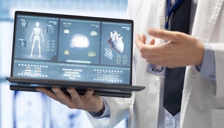
In addition to 3D imaging, advanced analysis techniques are revolutionizing the field of radiology. These technologies allow radiologists to analyze medical images in greater depth, providing more insights for diagnoses and treatment decisions.
AI and Deep Learning
One of the most exciting advancements in imaging analysis is the integration of artificial intelligence (AI) and deep learning algorithms. These technologies help analyze 3D X-ray images with higher accuracy and speed than traditional methods. AI can be trained to detect abnormalities such as tumors, fractures, or infections, making the analysis process faster and more reliable.Computer-Aided Detection (CAD)
CAD systems assist radiologists by highlighting suspicious areas in X-ray images that may require further investigation. These systems improve the accuracy of diagnosis by providing a second set of "eyes" to detect early-stage conditions that might be overlooked.Quantitative Imaging
Quantitative imaging techniques involve the use of advanced software to extract numerical data from medical images. For example, these methods can measure the size and volume of tumors, assess bone density, or calculate the amount of plaque in blood vessels. This detailed analysis is critical for tracking disease progression and evaluating the effectiveness of treatments.Image Fusion and Multi-Modality Imaging
Advanced analysis also involves combining images from different imaging modalities, such as CT scans, MRIs, and X-rays. By fusing these images, doctors can gain a comprehensive understanding of a patient’s condition, seeing both soft tissue and bone structures in a single, detailed image. This technique is particularly useful in complex cases where a combination of images provides a better diagnosis.Virtual Reality (VR) and Augmented Reality (AR)
VR and AR are also being integrated into the analysis of 3D X-ray images. In some cases, doctors can use VR to "walk through" 3D models of patient anatomy, while AR can overlay diagnostic data onto a physical environment. These tools enhance the precision of surgeries, guide interventions, and improve patient outcomes.
Benefits of 3D Imaging and Advanced Analysis:
Improved Accuracy: imaging provides a better and more accurate representation of the internal structures within the body, minimizing the scope for missed diagnosis.
Better Treatment Planning: With sophisticated analysis software, physicians can better plan treatment, leading to better patient outcomes.
Faster Diagnosis: With 3D imaging and AI-aided analysis, diagnosis is better in most cases where time is critical.
Personalized Medicine: With 3D models and sophisticated analysis, physicians can personalize treatment for specific patients, targeting interventions more accurately and effectively.
Minimized Radiation Exposure: 3D imaging sometimes eliminates the need for serial imaging tests, reducing a patient's exposure to radiation.
Challenges and Future Directions:
While 3D imaging and sophisticated analysis offer spectacular advantages, there are hurdles to be addressed:
Cost: High-end imaging and analysis technology such as 3D imaging comes at a high cost, which can function as a barrier to access in low-resource environments.
Training and Expertise: Clinicians and radiologists must be well trained to utilize 3D imaging and high-level analysis to the fullest. It takes time and labor.
Data Security:Owing to the volume of data produced by 3D imaging still rising, patient confidentiality and secure data storage are matters of concern.
Other than this, the future of 3D imaging and advanced analysis in X-ray technology is rosy. With more software development, compatibility with AI, and availability, such technologies can only get better, boosting diagnosis and treatment of most health conditions.


Reference Website Link:
National Institute of Biomedical Imaging and Bioengineering (NIBIB) - 3D Imaging in Medicine
Link: https://www.nibib.nih.gov/science-education/science-topics/3d-imaging
Radiological Society of North America (RSNA) - 3D Imaging in Radiology
Health Imaging - 3D Imaging in Radiology
Radiology Today - The Rise of 3D Imaging in X-ray
Link: https://www.radiologytoday.net/archive/rt1019p10.shtml
Journal of Medical Imaging - 3D Imaging Techniques and Applications
Innovative
Contact Us
Service
xraybazar.com
© 2024. All rights reserved.
Address- Rajasthan, India
Gmail Id- xraybazaroffcial.com
Important Links